Brain Mapper
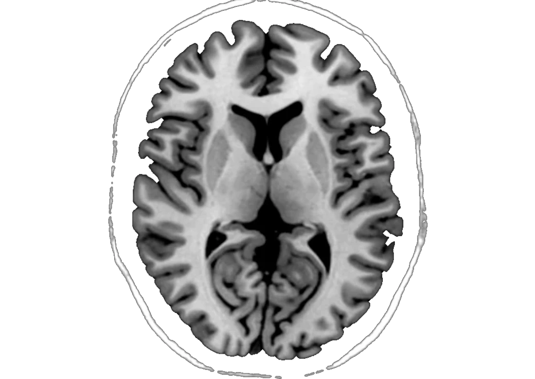
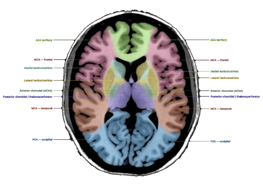
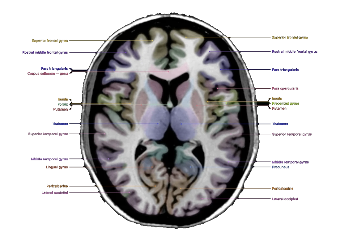
Every lobe, gyrus and deep nucleus mapped on your own 3-D T1 MRI, in three linked planes, in under a minute.
- Choosethe study folder. We find the 3-D T1.
- De-identifyon your computer: tags, text and face removed.
- Explorethe labelled brain with clinical notes.
Private, fast, and checked against expert tracings
What leaves your computer
Only the head volume, resampled to 1 mm, with the face and background removed. No DICOM header, file name or date is ever sent.
What we keep
The scan is deleted from our server as soon as it is mapped. The map is kept for 7 days so you can reopen it; you can delete it sooner.
Scans it accepts
3-D T1 MPRAGE, BRAVO or 3D TFE with thin slices, as uncompressed DICOM or NIfTI. One study at a time.
- Select the 3-D T1
You choose the study folder. Your browser reads every series and picks the thin-slice 3-D T1 (MPRAGE, BRAVO, 3D TFE). Nothing is uploaded at this stage.
- De-identify on your computer
All DICOM tags are dropped, burned-in text and background are cleared, and a landmark network running in your browser finds and removes the face. You review a preview before anything is sent.
- Segment
On our cloud GPU, a 3-D convolutional neural network trained by NeuroEvidence on NVIDIA A100 GPUs labels every voxel into 100 structures in about 42 seconds. The uploaded scan is deleted as soon as it is mapped.
- Explore and measure
The labelled brain opens in three linked planes with clinical notes for each structure. Optional volumetrics compare each structure with 1,027 healthy adults; the map and report can be downloaded.
Method
| Input | 3-D T1-weighted MRI with thin slices (about 1.5 mm or less), 1.5 T or 3 T, as DICOM or NIfTI. One study at a time. |
|---|---|
| Pre-processing in your browser | Series selection, removal of all DICOM metadata, clearing of burned-in text and background, face removal by our landmark network, then resampling to a 1 mm isotropic 256³ volume (trilinear) with intensity normalisation. |
| Network | 3-D U-Net style convolutional network, about 13 million parameters, designed and trained by NeuroEvidence. The head is processed in overlapping 128³ blocks whose class probabilities are combined with Gaussian weighting, so there are no seams between blocks. |
| Labels | 100 structures: 62 cortical regions (31 gyri per hemisphere, Desikan–Killiany–Tourville protocol), deep grey nuclei, hippocampi and amygdalae, ventricles and temporal horns, brainstem, cerebellum, corpus callosum, cerebral white matter and CSF. Each label is cleaned to its main connected piece. |
| Training | About 1,500 scans from seven public research collections (healthy adults and stroke patients, 1.5 T and 3 T), then a second stage on 71 brains whose cortex was traced by hand by neuroanatomists (Mindboggle-101). Random rotation, scaling, contrast and noise during training for robustness to unseen scanners. Trained on NVIDIA A100 GPUs. |
| Volumetrics optional | Head size estimated from the map and calibrated against a standard intracranial-volume method. Each structure is expressed as a percentile for age, sex and head size against 1,027 healthy adults aged 20–89 (IXI and Dallas Lifespan Brain Study, four scanners). Quality check: the scan is also mapped as its mirror image; structures where the two maps disagree are not scored. |
| Compute | Mapping runs on cloud servers on one NVIDIA L4 GPU and takes about 42 seconds per scan (about 1.5 minutes with the volumetrics report, which maps the scan twice); no installation or GPU is needed on your side. |
| Validation | Held-out brains never used in training, scored against expert hand tracings with the Dice overlap. See the Validation tab. |
- Labelling protocol: Klein A, Tourville J. 101 labeled brain images and a consistent human cortical labeling protocol. Front Neurosci 2012;6:171.
MRI scans
Seven public research collections from different hospitals and scanners, 1.5 T and 3 T: healthy adults from their late teens to their seventies, and stroke patients, so the network has seen brains with lesions.
Expert-traced brains
A second training stage on brains whose cortex was traced by hand by neuroanatomy experts, so the gyral boundaries follow expert anatomy rather than software defaults.
Parameters, trained by us
A 3-D convolutional network designed and trained by NeuroEvidence. During training every scan is randomly rotated, rescaled and given different contrast and noise, so it copes with scanners it has not seen.
Brains used to measure accuracy (Validation tab) were held out and never used in training.
Accuracy is the overlap between each tool’s labels and labels drawn by hand by experts (Dice score: 1.0 = identical). Every tool mapped the same held-out scans, none of which was used to train Brain Mapper.
| Tool | Cortex 20 brains, 62 regions | Deep nuclei 20 brains, 14 nuclei | Deep nuclei second test set, 10 brains | Time per scan | De-identification |
|---|---|---|---|---|---|
| Brain Mapper | 0.821 | 0.790 | 0.749 | about 42 s on one L4 GPU | ✓ built in, in your browser |
| FreeSurfer 8.1 (recon-all) | 0.813 | 0.789 | 0.743 | about 87 min on CPU (4 cores) | Not available |
| SynthSeg (FreeSurfer 8.1) | 0.649 | 0.779 | – | about 1.5 min on CPU (4 cores) | Not available |
Where Brain Mapper is ahead
- More accurate than FreeSurfer on the cortex in 95% of brains. Closer to the expert tracing in 19 of 20 brains; 0.821 vs 0.813, paired difference +0.007 (95% CI +0.005 to +0.010).
- Same accuracy on the deep nuclei, in a fraction of the time. 0.790 vs 0.789 on the expert tracings and 0.749 vs 0.743 on the second test set, in about 42 seconds instead of about an hour and a half.
- Nucleus accumbens, caudate and putamen. Accumbens 0.67 vs 0.63 and caudate 0.88 vs 0.86 against FreeSurfer on the expert tracings; putamen ahead on the second test set.
- Cortex vs SynthSeg. 0.821 vs 0.649 on the expert cortex tracings.
- Nothing to install. Runs in your web browser; the others are installed and run from the command line.
- Built-in de-identification. The face, DICOM tags and burned-in text are removed on your computer before upload.
- Made for teaching. Three linked planes with clinical notes for each structure, and a downloadable standalone version.
- Test sets: 20 OASIS-TRT-20 brains from Mindboggle-101 (expert cortex and deep-nucleus tracings) and 10 brains from the Hammers adult atlas (structures drawn from scratch). Mindboggle’s experts edited FreeSurfer starting labels, which slightly favours FreeSurfer.
- Where FreeSurfer is ahead: the globus pallidus (0.79 vs 0.86 on the expert tracings) and, on the Hammers atlas, the ventricles and temporal horns (0.76 vs 0.77 overall). SynthSeg is designed to work on any MRI contrast; its cortex is coarser on standard T1.
- Scores are for Brain Mapper version 1.0 (November 2026). These test sets were also used during development; a confirmation on new test sets is planned. Times are for the mapping step on the hardware shown.
Brain Mapper is an educational and research tool. It is not a medical device and is not for diagnosis or treatment. Labels are automatic and can be wrong, especially near lesions. © 2026 NeuroEvidence.ai · About Labs & copyright