Angio Mapper
Every artery on a cerebral angiogram outlined in seconds, from the carotid siphon to the small cortical branches.
- Dropa DSA image: PNG, JPEG or a screenshot.
- Mapon our server in seconds. Nothing is stored.
- Explorethe vessel map over the image.
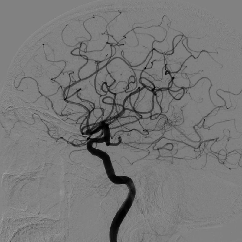
 BeforeAfter
Works on subtracted catheter angiograms (DSA), frontal or lateral, arterial phase. Vessels may be dark or white. CT and MR angiograms are not supported yet.
Private, fast, and trained by us
Mapped on our server, never stored
Your image is sent to our server, mapped, and deleted as soon as the map comes back. It is never saved, logged or used for training.
De-identification
We recommend using only images with no patient information. As an extra layer of protection, burned-in names, dates and numbers are detected and removed in your browser before the image is sent, with a warning.
What it accepts
A single DSA frame as PNG, JPEG or WebP, at least about 400 pixels across. Pick a late arterial frame for the fullest map.
Angiograms first
Stage 1: X-ray angiograms with traced vessels, so the network learns what a contrast-filled vessel looks like before it sees the brain.
Acute stroke angiograms
Stage 2: 30 cerebral DSA frames from stroke patients, frontal and lateral, with expert vessel tracings. 10 more frames chose the best checkpoint. The 20 test frames were never used for training or any choice.
Parameters, trained by us
A 2-D U-Net designed and trained by NeuroEvidence. Each training image is randomly rotated, rescaled, flipped, inverted and given different contrast, blur, noise and resolution, so it copes with other machines and screenshots.
| Setting | Value |
|---|---|
| Network | 2-D U-Net, 4 down-sampling steps, 24 to 384 channels, group normalisation, SiLU; 4,367,641 parameters |
| Input | One grey-scale frame, intensities scaled between the 1st and 99th percentile; any size (trained on 512 x 512 crops) |
| Loss | Binary cross-entropy + soft Dice. Stage 1: background weight 0.15 where only some vessels are traced |
| Optimiser | AdamW, learning rate 2e-3 (stage 1) then 1e-3 (stage 2), batch 12, weight decay 1e-4 |
| Checkpoint | Best Dice on the 10 validation frames (0.785), checked every 250 steps |
| Running it | ONNX Runtime on our server; map shown at 0.2 by default (Sensitivity slider; the validation scores use 0.5); loose pieces under 25 pixels removed |
Each model mapped the same 20 hand-labelled angiograms from 20 patients that Angio Mapper never saw, and was scored against the expert tracing of each frame. Scores are the mean over the 20 images, with the 95% confidence interval (bootstrap) for Dice. 1.0 = identical to the expert.
| Model | Dice overlap, 95% CI | clDice vessel tree kept connected | Sensitivity expert vessel found | Precision mapped pixels that are vessel | ROC AUC |
|---|---|---|---|---|---|
| Angio Mapper | 0.763 0.746 to 0.778 | 0.706 | 0.783 | 0.753 | 0.977 |
| VSS-Net, single frame (published, same 20 images) | 0.760 | – | – | – | – |
| Frangi vessel filter (classical, tuned on the validation frames) | 0.489 0.440 to 0.538 | 0.523 | 0.448 | 0.558 | 0.859 |
What the numbers show
- Matches the published single-frame result. 0.763 vs 0.760 for VSS-Net, the published single-frame network, on the same test images.
- Far ahead of a classical vessel filter. Better on all 20 test images (Dice +0.27, clDice +0.18; Wilcoxon p < 0.001).
- Large and medium vessels are found reliably. 98% of large (8 px or wider) and 94% of medium (4 to 8 px) expert centreline is mapped.
- Robust to format. Dice 0.754 with the image inverted (white vessels) and 0.759 at half resolution, the common changes in screenshots and exports.
- Consistent. Every test image scored between 0.68 and 0.83; none failed.
Where it is weaker
- The thinnest branches. Only 53% of expert centreline narrower than 4 px is mapped. Most disagreement with the expert is here.
- One dataset. All test frames come from one stroke cohort with one expert tracing per frame, so agreement between two experts could not be measured. An external test on a second dataset is planned.
- Removing loose pieces under 25 pixels is a display choice; it changes test Dice by less than 0.001.
Test set: 20 hand-labelled angiograms. Sources and references are on the copyright page. Researchers who want the trained model or the evaluation script for non-commercial research can contact us.
- Name the arteries
Label each vessel on the map: ICA, M1, M2, ACA, PCA, basilar and their branches.
- Flag what looks abnormal
Teaching cases for cut-offs and aneurysms, built with expert review.
Angio Mapper is an educational and research tool. It is not a medical device and is not for diagnosis or treatment. The map is automatic and can miss or add vessels. © 2026 NeuroEvidence.ai · Copyright